Looking for Something?
Location & Hours
719 W Main St
Atlanta, TX 75551
| M, W, Th, F: | 8:00am - 5:00pm |
| Tuesday: | 8:00am - 6:00pm |
Related Articles
- 10 Eye Jokes Just for You!
- 10 Eye Related Jokes to Brighten Your Day
- 10 Fascinating Facts about the Eye
- 10 Fun and Fascinating Eye Facts
- 10 Super Cornea Optical Jokes Just for You!
- 10% of Glaucoma Cases are Closed or Narrow Angle
- 11 Bad Contact Lens Habits
- 11 Fun and Fascinating Eye Facts
- 12 Terrible Eye Jokes
- 13 MORE Eye Jokes Just for You!
- 13 More Eye Related Jokes to Brighten Your Day
- 13 More Super Cornea Optical Jokes Just for You!
- 20-20-20 Rule for Better Vision
- 2019 Most Dangerous Toys for Kids
- 2020 Most Dangerous Toys
- 4 Good Reasons for Older Adults to Have Regular Eye Exams
- 4 Reasons Your Eyelid Might Be Twitching
- 5 reasons not to buy your eyeglasses online
- 5 Reasons to Avoid Internet Eyeglasses
- 5 Reasons to Buy Glasses Locally
- 5 Reasons to Buy Your Glasses Locally
- 5 Ways to Take Care of Your Optical Assets
- 6 Activities That Can Change Your Eye Pressure
- 6 Reasons Sunglasses Are Essential
- 6 Reasons Sunglasses Are Essential
- 7 Tips For Getting The Most Out Of Your Eye Exam.
- 7 Tips for Your Best Eye Exam
- 7 Tips for Your Next Eye Exam
- 7 Tips from an Eye Doctor on Getting the Most from Your Exam
- 9 Fascinating Facts About Green Eyes
- 90% of Glaucoma Cases are Open Angle
- A Child's Vision
- Activities That Can Change Your Eye Pressure
- ADHD and Your Eyes
- Alcon DAILIES TOTAL1
- Allergies are one of the most common eye conditions
- Am I Dealing with Allergies, Dry Eye, or Infection?
- Annual Eye Exams-Good for Your Health and Your Wallet
- Anti-Reflective Lenses
- Are My Cosmetics Making My Dry Eye Worse
- Are My Eyes Changing Colors?
- Are My Meds Making My Eyes Drier?
- Are My Reading Glasses Making My Eye Worse?
- Are Reading Glasses Making My Eyes Worse?
- Are You Asking for Trouble with Your Contacts Care Routine?
- Astigmatism
- Astigmatism is NOT a Scary Diagnosis
- Back-to-School Eye Exams
- Bargain Halloween Contacts?
- Basics of Macular Degeneration
- Best Place to Turn for Help for a Red Eye
- Bilberry & Macular Degeneration
- Blepharitis
- Blepharitis 101
- Bring Your Glasses to the Eye Doc...Even if You HATE Them!
- Can Drug For Rheumatoid Arthritis Damage Eyes?
- Can I Have Cataract Surgery if I Have Macular Degeneration?
- Can you guess the most dangerous sports for eye injuries?
- Can't Learn to Live with Those Eye Floaters?
- CareCredit - Benefits Are All Around
- Carrera 85 Icons
- Cataract Surgery
- Cataract Surgery and Anesthesia Types
- Cataract Surgery and Life Expectancy
- Cataract Surgery? I see fine!
- Cataracts
- Choosing The Right Eyeglass Frames For Your Face
- Computer Vision Glasses
- Computer Vision Syndrome
- Contact Lens Care 101
- Contact Lens Hygiene 101
- Corneal Molding or Ortho-K
- Coronavirus and Your Eyes
- Costume Contacts Can Make Halloween a Scary Time
- Dealing with Macular Degeneration
- Dealing with Macular Degeneration
- Demodex--the Mite that Lives Among Your Eyelashes
- Demodex: The Weird Little Mite Living in Your Eyelashes
- Detecting Alzheimer's Disease through the Eyes?
- Detecting Alzheimer's though an Eye Exam?
- Diabetic Retinopathy
- Diabetic Retinopathy Basics
- Diabetic Retinopathy Diagnosis and Treatment
- Diabetic Retinopathy Explained
- Diabetic Retinopathy Must-Knows for Everyone with Diabetes
- Diabetic Retinopathy--Diagnosis & Treatment
- Diagnosis and Treatment of Diabetic Retinopathy
- Did You Know - Age-Related Macular Degeneration
- Did You Know You Can Get Freckles in Your Eye?
- Differing Functions of Ophthalmologists, Optometrists, and Opticians
- Dilation Exam
- Do Erectile Dysfunction (ED) Drugs Really Cause Vision Loss?
- Do I Even Need an Exam?
- Do I Have Eye Allergies? If So, What Can I Do?
- Do I Have the Dreaded Pink Eye?
- Do I Really Need Cataract Surgery?
- Does Your Child Have Undetected Vision Issues?
- Don't Let Your Child's Vision Issue Go Undetected
- Don't Play Trick or Treat with Your Eyes
- Dropless Cataract Surgery
- Dropless Cataract Surgery Is Now Available
- Drug Allergy or Side Effect? Knowing the difference could save your life
- Dry Eye Risk Factors
- Dry Eye Syndrome
- Dry Eyes or Allergies? Or Something Else?
- Dry Eyes Or Allergies? Which Do I Have?
- Dry Eyes Or Allergies? Which Do I Have?
- Eat Your Way to Better Eye Health
- Eating Your Way to Better Eye Health
- Emergency Room Not Usually Best Choice for Red Eye
- Every Pink Eye Is NOT 'Pink Eye'
- Explosion in Fireworks Eye Injuries
- Eye Allergies and You
- Eye Anatomy Crossword Puzzle
- Eye Color and You
- Eye Jokes--Our Gift to You!
- Eye Jokes--Our Holiday Gift to You!
- Eye Liner, Shadow, and Your Dry Eyes
- Eye Problems Associated with Lyme Disease
- Eye Safety During the Holidays
- Eye Safety for the Solar Eclipse
- Eye Safety for the Upcoming Solar Eclipse
- Eye Safety on the 4th of July
- Eye Safety: The Worst Toys for Kids
- Eyes feel Like the Sahara Desert : What do I do?
- Familiar with the 20-20-20 Rule?
- Flashes and Floaters
- Floater Relief with Lasers
- Geriatric Eyes
- Geriatric Vision
- Giving the Gift of Sight
- Glasses=Need, Contacts=Luxury.
- Glaucoma
- Glaucoma - Open Angle and Normal Tension
- Glaucoma & Sleep Apnea
- Glaucoma Narrow Angle
- Glaucoma: Occupational Hazard for Musicians?
- Had LASIK? Get a Copy of Your Eye Records ASAP!
- Halloween Hazards
- Handling your contact lenses
- Happy 2020!
- Health or Vision Insurance?
- Health vs Vision Insurance
- Hearing That Your Driving Days Are Over
- Help for Those with Low Vision
- Help Your Child See Their Way to a Better School Year
- Help! Is My Eye Bleeding?
- Help! Growing Older and Can No Longer Read with My Contacts!
- Help! I started seeing these floating things!
- Hereditary Eye Diseases
- Holiday Eye Safety
- Holiday Eye Safety Tips
- How Many of These Bad Contact Lens Habits Are You Guilty Of?
- How Parkinson’s Disease Affects the Eye
- How Shingles Can Affect Your Eye
- How to Deal with Your Scary Red Eye
- How to Give the Gift of Sight
- How to insert contact lenses
- How to Make Your Red Eyes Redder
- How to Make Your Red Eyes Worse
- How to remove contact lenses
- How to Ruin a Fun Day in the Water in One Easy Step
- How We Test Your Eye Pressure
- Hyperopia Myopia
- I Have Macular Degeneration - Should I Take Bilberry?
- I Should Pay Out-of-Pocket for Cataract Surgery Now?
- I Think My Reading Glasses Made My Eyes Worse!
- I'm 45 and My Contacts No Longer Work for Me!
- I'm seeing jagged lines in my vision! Help!
- If You've Had LASIK, Get Your Eye Records NOW!
- In a Flash...Your Eyes & Fireworks Injuries
- Intraocular lens (IOL)
- Intraoperative Aberrometry & Cataract Lens Replacement Selection
- Is an Eye Exam on Your Back-to-School Checklist?
- Is Coffee Bad for My Eyes?
- Is Coffee Good or Bad for My Eyes?
- Is It a Drug Allergy or a Side Effect?
- Is My Coffee Addiction Bad for My Eyes?
- Is My Eye Bleeding?
- Is My Eye Makeup Making My Dry Eyes Worse?
- Is There Anything I Can do to Stop Getting More Nearsighted?
- Just Found Out I Have Primary Open Angle Glaucoma. What Now?
- Keeping Your Glasses in Working Order
- Keratoconus
- Know the 3 F's of Retinal Detachments
- Laser Treatments Might Help Floaters
- LASIK
- Love Is in the Air, Love Is in the Eyes
- Love Is in the Air...or Eyes
- Lyme Disease And Your Eyes
- Macular Degeneration
- Macular Degeneration & Bilberry
- Macular Degeneration 101
- Macular Degeneration--What Can Be Done?
- Macular Degeneration, Cataracts, and You
- Macular Mojito and Cataract Cocktails?
- Marco - Redefining the art of refraction
- Maui Jim - Like You Have Never Seen
- Maui Jim Commercial 2016
- Maui Jim Lens Technology
- Minimally Invasive Glaucoma Surgery
- Mom's Eyes
- Mucus Fishing Syndrome
- My Eyeballs Can Get Wrinkles, Too?
- My Eyes Feel Like a Desert!
- My Eyes Feel Like the Sahara!
- My New Glasses Are Too Strong!
- My New Glasses Aren't Working for Me
- New Transition Contact Lenses
- Newly Diagnosed with Glaucoma? What Now?
- Ophthalmologists, Optometrists, Opticians - What's the Difference?
- Optical Jokes--Our Holiday Gift to You!
- Optical Jokes: Our Gift to You!
- Options for Vision Correction While Playing Sports
- Parkinson's & Your Eyes
- Polarized Sunglasses
- Pregnancy and Your Eyes
- Pregnancy Can Change Your Eyes
- Presbyopia
- Primary Open Angle Glaucoma
- Progressive Lenses
- Punctual Plugs
- Refractive Error
- Retinal Detachment - Symptoms, Causes, and Treatment
- Retinopathy
- Rheumatoid Arthritis, Hydroxychloroquine, and Your Eyes
- Science and a Mother's Eyes
- Screen Time & Your Eyes
- Seeing one of the 3 F’s
- Shingles and Your Eye
- Should I Be Scared of My Astigmatism Diagnosis?
- Should I Worry About Astigmatism?
- Should These Flashes & Floaters Worry Me?
- Silhouette of the Time
- Sleep Apnea & Glaucoma: There's a Link
- Slit Lamp
- Special Ops Eyes, the Vision of our Military
- Sports and Eye Injuries
- Sports and Vision Correction
- STOP Fishing Mucous out of Your Eyes!
- Sunglasses in the Winter?
- Suspect ADHD? Time for an Eye Exam!
- Swimming and Contacts Don't Mix
- Taking Care of Your Child's Eyes
- Texting and Driving = Bad Idea
- Texting and Driving? Not a Good Combo.
- The 3 F's--and Why They Can Be So Dangerous
- The Benefits of Daily Disposables
- The Care & Cleaning of Your Glasses
- The Eyes You are Born With Are the Eyes You Keep - AOA
- The Hallmark Sign of Ocular Allergies
- The Importance of Knowing if it Was a Side Effect or a True Allergy
- The Importance of Polarized Eye Wear During The Winter
- The Importance of Quality Sunglasses
- The Leading Cause of Blindness in Working-Age Adults
- The Link Between Sleep Apnea and Glaucoma
- The Lowdown on Low Vision
- The Perfect Frame for You!
- The Power of a Mother's Eyes
- The Scary Cost of Cheap Costume Lenses
- The Three O's of Eye Care: Ophthalmologist, Optometrist and Optician
- The Wonderful World of Dailies
- The World of Eye Color
- There's Blood in the Back of My Eye?!
- These weird spots and jagged lines are scaring me!
- Things That Make Your Eye Twitch
- Think of Them as Sunscreen for Your Eyes
- Through a Mother’s Eyes
- Through a Mother’s Eyes
- Time For Some Holiday Eye Care Jokes
- Time to Hang Up the Keys?
- Time to Hang Up the Keys?
- Tips for Making Those New Glasses Last
- Tips to Save Your Eyes This Holiday Season
- Top 4 Reasons Every Older Adult Needs Regular Eye Exams
- Top 4 Reasons You Need Your Eyes Checked More Frequently as You Get Older
- Top 6 Reasons To Wear Sunglasses
- Transitions Contact Lenses
- Try saying Intraoperative Aberrometry 3 Times Fast!
- Two Common Genetic Eye Diseases
- Two Common Genetic Eye Diseases
- Ultra-Violet Light
- UV Damage in Childhood
- Vision Hallucinations and Charles Bonnet Syndrome
- Vision or Health Insurance?
- Vision Problems After a Concussion or TBI?
- Vision Problems Can Happen After A Concussion
- Vision Problems Following Concussion
- Visit our new website!
- Visual Hallucinations? It Could Be Charles Bonnet Syndrome.
- Want to Know How to Make Your Red Eyes Redder?
- Want To See Better? Live a Healthy Lifestyle
- Ways Eyes Can Change During Pregnancy
- What 20/20 Vision Actually Means
- What About Cataract Surgery with My Macular Degeneration?
- What Anesthesia Might You Have for Cataract Surgery?
- What Are Retina Wrinkles?
- What are the risk factors for dry eye?
- What Are These Floating Things?
- What can I do for my red and itchy eyelids?
- What does ‘20/20 vision’ actually mean?
- What Everyone Should Know About Macular Degeneration
- What Happened To Reversal Drops For Eye Dilation
- What Happened to Those Dilation Reversal Drops?
- What Is 20/20 Vision?
- What is a "Progressive" lens and do I need one?
- What is a freckle in the back of my eye?
- What Is a Punctal Plug and Why Would I Need One for My Eyes?
- What Is a Punctal Plug?
- What Is a Retinal Vein Occlusion?
- What is Diabetic Retinopathy?
- What Is Low Vision and How Is It Treated?
- What is Minimally Invasive Glaucoma Surgery?
- What Is Mucus Fishing Syndrome?
- What is POAG, or Primary Open Angle Glaucoma?
- What Is Refraction & Why Doesn't Health Insurance Cover It?
- What Is Refraction & Why Doesn't My Health Insurance Cover It?
- What Is Refraction And Why Doesn’t Insurance Always Cover It?
- What Is the Deal with Those Weird Cataract Glasses?
- What Is This Bump on My Eyelid?
- What Kind of Eye Correction Do I Need for Sports?
- What makes a 'Progressive' Lens Progressive?
- What Sunglasses Can Do for You
- What Testing Might I Have During My Eye Exam?
- What Tests Might I Have During My Eye Exam?
- What Tests Might You Have During Your Eye Exam?
- What to Do About Those Pesky Floaters
- What To Expect During An Eye Injection
- What to Know Before Your First Eye Injection
- What to Know Before Your First Eye Injection
- What You Should Know About Intraoperative Aberrometry Before Your Cataract Surgery
- What You Should Know About MIGS (Minimally Invasive Glaucoma Surgery)
- What's a retinal detachment and what are the symptoms?
- What's that lump on my eyelid?
- When Do You NEED Cataract Surgery?
- When Should I Stop Driving?
- When should my child get an eye exam
- When Your Retina Gets a Wrinkle
- Which Eye Diseases Run In Families?
- Why a Progressive Lens Might Be Your Best Choice
- Why am I having Difficulty Adjusting to My New Glasses?
- Why Are My Eyes Changing Color?
- Why Are My Eyes Red?
- Why Are They Saying I Need to Pay Out-of-Pocket for Cataract Surgery?
- Why Dilation Reversal Drops Are Not Currently an Option
- Why Do I have to Pay Out of Pocket for Cataract Surgery?
- Why Do I Need an Eye Exam When I Can See Great?
- Why Do I Need an Injection in My Eye?
- Why Do I Need an Injection in My Eye? Part 2
- Why do I need glasses if I have contact lenses?
- Why Do People Wear Those Big Sunglasses After Cataract Surgery?
- Why Do You Have to Touch My Eye or Puff It with Air?
- Why everyone who has had LASIK surgery should get their records NOW
- Why Frequent Eye Exams Are a MUST If You Take Hydroxychloroquine (Plaquenil)
- Why Get an Early Eye Exam for Your Young Child?
- Why Good Sunglasses are a Great Investment
- Why Having Astigmatism Isn't Usually That Big a Deal
- Why is my eye twitching?
- Why is there Blood in the Back of My Eye?
- Why Polarized Glasses Are Essential in Winter
- Why Rubbing Your Eyes Is a Mistake
- Why Should I Know What Intraoperative Aberrometry Is?
- Why Those Cheap Halloween Costume Lenses Aren't Worth the Cost
- Why You Need to Bring Your Current Glasses Even if You Hate Them
- Why You Shouldn't Try to Get By with Just Contact Lenses
- Why Your Child NEEDS an Eye Exam
- Wiley X Survival Test - Hammer
- Wiley X Survival Test - Shotgun
- Winter Is Prime Time for Polarized Sunglasses!
- You Want to Do WHAT to My Eye? Part 1
- You Want to Do WHAT to My Eye? Part 2
- You Want to Inject My Eye? Part 1
- You Want to Inject My Eye? Part 2
- Your Child & Your Eye Doctor
- Your Child's Eyes & ADHD
- Your Eye & Shingles
- Your Eyes + Lyme Disease
- Your Meds = Dry Eyes?
- Your Vision & Parkinson's Disease
Related Tags
Blog
The colored part of the eye is called the iris. It is composed of two muscles. The constrictor and the dilator. The black circle at the center of the iris is called the pupil and is actually just an opening to allow light into the eye. The pupil appears black because the inside of the eye contains pigment to absorb all of the light that enters it, not allowing any of it to reflect back out.
When light enters your eye there is a muscular reflex in your iris to regulate the amount of light that hits the retina. Too much light and constrict muscles tighten to making your pupil smaller to limit the amount of light entering your eye. Not enough light and the dilator muscles contract allowing your pupil to get bigger.
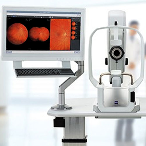
During a comprehension eye exam, your eye doctor might want to dilate your pupils to evaluate the back of each eye under high magnification. Without dilation, the bright exam light automatically causes your pupils to constrict, making a much smaller opening to look through.
Dilating drops cause your pupils to enlarge by inhibiting the constrictor muscles from reacting to light., then the doctor can use a bright exam light combine with a magnification lens for an unobstructed view of the internal structure of each eye, including the retina, macula and optic discs.
Dilation drops typically cause blurred near-vision and light sensitivity lasting about 4 to 6 hours. Sunglasses are usually worn afterward to relieve glare discomfort, but you should be safe to drive with caution.
Keratoconus is an eye disease in which the cornea deforms from its normally curved dome shaped and becomes cone shaped. Sometimes there is a flaw in the collagen, the material of the cornea that weakens and allows the cornea to stretch into an irregular cone shape.
The cornea is the clear tissue located at the front of the eye and it refracts and focuses light as it enters the eye. Therefore abnormalities of the corneal surfaces can severely distort vision.
Symptoms usually start in the teen years with near-sighteness and astigmatism which can often be treated with contact lenses or glasses. At the onset it can be difficult to detect.
It is first diagnosed when the cornea starts reveal progressive irregular distortion and eventually becomes to advance for conventional glasses or contact lenses to correct. At this critical point it is often treated with specially designed contact lenses to provide a smooth optical surface to focus light rays and impede progression of the bulging cone shape.
In some case keratoconus can progress to the point that corneal replacement surgery is needed. This usually occurs around the age of 35.
There are some evidence to suggest allergy suffers and people with rigid contact lenses that rub their eyes may contribute to the progression of the symptoms and cause scratches on the surface of the cornea.
If you feel you are at risk or your prescription is changing rapidly make certain your eye care professional checks for Keratoconus during your next eye health and visual examination.